Ver ítem
- xmlui.general.dspace_homeCentros Regionales y EEAsCentro Regional Buenos Aires SurEEA BalcarceArtículos científicosxmlui.ArtifactBrowser.ItemViewer.trail
- Inicio
- Centros Regionales y EEAs
- Centro Regional Buenos Aires Sur
- EEA Balcarce
- Artículos científicos
- Ver ítem
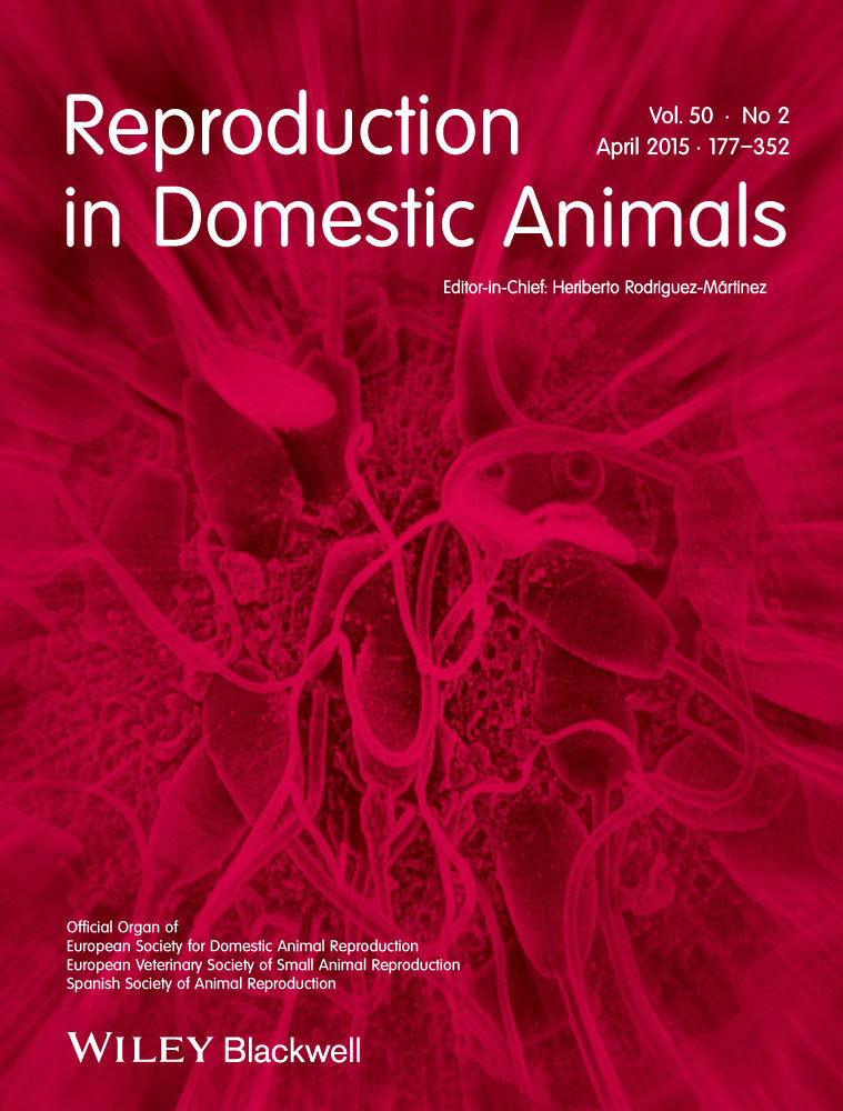
Vaginal histological changes after using intravaginal sponges for oestrous synchronization in anoestrous ewes
Resumen
To characterize the histological and cytological vaginal changes generated by the use of intravaginal sponge (IS) applied in oestrous synchronization treatments in ewes during mid‐non‐breeding season. Thirty‐five multiparous ewes were allocated to three experimental groups according to the moment in which the samples were taken: (i) ewes treated with IS containing 60 mg of medroxyprogesterone acetate for 14 days, sampled the day of IS removal (group ISR;
[ver mas...]
To characterize the histological and cytological vaginal changes generated by the use of intravaginal sponge (IS) applied in oestrous synchronization treatments in ewes during mid‐non‐breeding season. Thirty‐five multiparous ewes were allocated to three experimental groups according to the moment in which the samples were taken: (i) ewes treated with IS containing 60 mg of medroxyprogesterone acetate for 14 days, sampled the day of IS removal (group ISR; n = 10), (ii) or after sponge removal at time of oestrus or 72 h after removal (group AR; n = 14) and (iii) ewes without sponge treatment that were sampled at the day of IS removal of the other groups (group CG; n = 11). Vaginal biopsies and cytological samples were taken from the anterior vaginal fornix area. The vagina of the CG group had a stratified squamous epithelium with a moderate degree of cellular infiltration with lymphocytes and plasma cells in the lamina propia. Treated ewes (ISR and AR) had epithelial hyperplasia and hypertrophy. ISR ewes had haemorrhage and perivascular infiltrate and an increased number of epithelial cells, neutrophils, macrophages and erythrocytes at IS removal. The use of IS generated histological and cytological alterations in the vaginal wall when used for oestrous synchronization in anoestrous ewes.
[Cerrar]

Autor
Manes, Jorgelina;
Campero, Carlos Manuel;
Hozbor, Federico Andres;
Alberio, Ricardo;
Ungerfeld, Rodolfo;
Fuente
Reproduction in Domestic Animals 50 (2) : 270-274 (April 2015)
Fecha
2015-04
Editorial
Wiley
ISSN
0936-6768
1439-0531
1439-0531
Formato
pdf
Tipo de documento
artículo
Palabras Claves
Derechos de acceso
Restringido
 Excepto donde se diga explicitamente, este item se publica bajo la siguiente descripción: Creative Commons Attribution-NonCommercial-ShareAlike 2.5 Unported (CC BY-NC-SA 2.5)

